Posts

Showing posts from July, 2021

DENTAL WAX-TYPES,INLAY WAX IN DETAIL NOTES

Image
  DENTAL WAXES  Low molecular weight ester of fatty acids derived from natural or synthetic components such as petroleum derivatives that soften to a plastic state at relatively low temperature  Wax has been a valuable commodity for over 2000 years  Carnauba wax:hardest,more durable  CLASSIFICATION OF WAXES: 1)ACCORDING TO ORIGIN: NATURAL : MINERAL :Paraffin,ceresin Carnauba,candelilla PLANT;Carnauba,candelilla INSECT:Beeswax ANIMAL:Spermaceti wax SYNTHETIC WAX Acra wax Aldo wax 2)ACCORDING TO USE AND APPLICATION: A)PATTERN WAXES  Inlay wax Casting wax Base plate wax B)PROCESSING WAX: Boxing wax utility wax Sticky wax C)IMPRESSION WAX Bite registration or corrective wax BOXING WAX Mode of supply : sheets  Uses:controls the thickness of the border ,preserves the extension UTILITY WAX: Composition:beeswax,petrolatum Mode of supply :sticks and sheets  Properties:soft at room temperature  Uses:to build up border of impression tray attaching a pontic of a bridge STICKY WAX Composition:yello

Leptospirosis -cause,pathogenesis,clinical features,diagnosis and treatment

Image
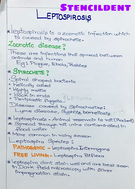
  LEPTOSPIROSIS  Leptospirosis is a zoonotic infection which is  caused by spirochete  ZOONOTIC DISEASE ? Zoonotic disease are infection that spread between animals and human  Examples :plague,ebola,rabies SPIROCHETE? Spiral shaped bacteria Helically coiled  Highly motile  Disease caused by spirochaetes: lyme disease,syphilis,borreliosis LEPTOSPIROSIS  Hook in ends Periplastic flagella  Leptospirosis -animal reservoir is rat (rodent ) Spread through rat urine contaminated in flood water More common in rainy season  LEPTOSPIRA SPECIES : PATHOGENIC -LEPTOSPIRA INTERROGANS FREE LIVING-LEPTOSPIRA BIFLEXA Leptospira dont stain well and are best seen in dark field microscopy with silver impregnation stain  PATHOGENESIS : Leptospira common in rodent (rat) ,also known as rat fever When a person who has cut or abraded skin walks in flood , Sewage water mixed with rat urine ;leptospira enter in and cause infection  Undergoes 2 phases: Phase 1- leptospiremia Phase 2-immune phase  Biphasic illness

Periodontal Screening And Recording -Periodontology easy notes

Image
 PERIODONTAL SCREENING AND  RECORDING  It has been developed jointly by the american academy of periodontology and the american dental association with the support of the procter and gamble company PROBE : WHO probe is used  It 0.5mm ball tip and colour coded from 3.5 to 5.5mm CODING SYSTEM : CODE 0 :  The probes coloured band remains completely visible  healthy and does not bleed on gentle probing  CODE 1:  Colour band completely visible in the deeper sulcus of the sextant bleeding after gentle probing is detected  CODE 2: Colour band completely visible,subgingival calculus defective margin are seen bleeding is there on probing  Treatment: Plaque and calculus removal,correction of plaque -retention margin of restoration and oral hygiene  CODE 3: Color band is partially visible  If 2 or more sextant score code 3,a comprehensive full -mouth examination and charting and indicated (3-4mm) CODE 4: Color band completely disappear (75mm) Asterisk* Abnormalities-furcation involvement ,tooth m

Examination of periodontium,periodontal pocket,teeth and implant

Image
  PERIODONTAL EXAMINATION OF  TEETH,IMPLANTS,PERIODONTIUM AND  PERIODONTAL POCKET  1)EXAMINATION OF TEETH AND IMPLANTS: A)TOOTH MOBILITY : All teeth have a slight degree of physiologic mobility which varies for : Different teeth Time  During the morning it will be greatest ,slight extrusion of tooth whereas ,during sleep it will be limited as there is not much  Occlusal contact these 24 hour variants are seen in those who  Have occlusal habits  such as : Bruxism Clenching LOCAL CAUSES: Periapical pathology Tooth morphology Implant mobility SYSTEMIC CAUSES : Age  Sex(females more predilection) Oral contraceptive Other Cause : Loss of tooth support Trauma from occlusion STAGES : 1)INITIAL /INTRASOCKET STAGE : Tooth moves within the confine of periodontal ligament  Associated with : Viscoelastic distortion Redistribution of periodontal fluid  2)SECONDARY STAGE: Occur gradually  Increase in horizontal force results in elastic deformation of alveolar bone  GRADING: According to ease and ext

Dermatophyte-What Is It ,Clinical Types,Lab Diagnosis And Treatment

Image
  DERMATOPHYTE  Infection caused by fungi is known as mycosis  Types: Superficial Subcutaneous Systemic SUPERFICIAL : Affect surface  Types : Superficial infection  Example -Tinea nigra,Piedra Cutaneous :Dermatophyte  DERMATOPHYTE : Superficial mycoses that affect only superficial keratinised tissue (hair,skin,nail) Break down and utilise keratin Infection caused it DERMATOPHYTOSES,tinea or ringworm GENUS ORDER  TRICHOPHYTON-Hair,skin,nail MICROSPORUM-hair,skin EPIDERMOPHYTON-skin,nail CLINICAL TYPES: Named based on site of involvement  Scalp-tinea capitis Groin-tinea cruris No hair involvement -tinea corporis Foot-tinea pedis or athlete's foot Beard -tinea barbae or barber's itch Chronic type with hair involvement is called FAVUS  FAVUS is followed by alopecia and scaring Its of two types ectothrix,endothrix Atherospore sheath over the hair shaft-ectothrix Atherospore within hair shaft -endothrix LAB DIAGNOSIS : SPECIMEN : Hair scraping,skin scraping ,nail DIRECT MICROSCOPY  1

Malignant Skin Tumors-Presentation,Clinical features,Management

Image
  MALIGNANT SKIN TUMORS  1)INTRODUCTION: Skin is outermost coat of human body  Skin act as a protective cover against various insulting agents such as ultraviolet radiation of sunlight 3 malignant skin tumour : Squamous cell carcinoma Basal cell carcinoma Malignant melanoma 2)PRESENTATION : A)SQUAMOUS CELL CARCINOMA: Carcinoma of cell of epidermis ,starts from prickle cell layer and migrate towards the outer surface. May occur in pre-existing lesion of skin: Marjolin ulcer Venous ulcer Leukoplakia Site: Junction of skin and mucous membrane eg(lip,eyelid) It is also seen in mucous membrane lined by stratified squamous epithelium (eg)tongue,mouth Predisposing factors: Scar,ulcer Exposure to sunlight or irradiation Bowen's disease Types: Proliferative Ulcerative  B)BASAL CELL CARCINOMA (RODENT ULCER) Common malignant skin tumors  Slow growing neoplasm Location: Majority of lesion found on face above a line from lobule of ear to angle of mouth Common Site : Eyelid Bridge of the nose Ar