Posts

Showing posts with the label oral medicine and radiology

Exposure and processing errors along with correction strategies -faulty radiograph part 2

Image
      FAULTY RADIOGRAPH -PART2                                     EXPOSURE ERROR: STUDENTS CORNER : Hi my dear stencilent family ,sorry for interepting, before moving on to the topic of discussion for today ,we also need to learn more reasons for faulty radiograpgh to occur along with its correction strategies to learn them , do click on the link provided  patient and positioning errors . DARK RADIOGRAPH: REASONS: PROLONGED EXPOSURE TIME (kvp) HIGH KILOWOLTAGE POTENTIAL(MA) Decreased film to source distance CORRECTION: Set exposure time kvp,MAcorrectly Adjust film to source distance LIGHT RADIOGRAPH REASONS : Low kvp -less MA Less exposure  Increased in film to source distance CORRECTION: Set exposure time kvp,MAcorrectly Adjust film to source distance PROCESSING ERROR: DARK RADIOGRAPGH : REASONS: Developing time to long High temperature of developer Inadequate fixation Light exposure before processing CORRECTION: Time -temperature method  68F-5 minute 78F-4 and half munute  LIGHT RAD

Patient and film positioning errors along with correction strategies - Faulty radiograph part 1

Image
             FAULTY RADIOGRAPH ARTIFACT : Unwanted density on the radiograpgh that interfere diagnosis. I)PATIENT AND FILM POSITIONING ERROR: FORESHORTENING: Reasons : increased vertical angulation  correction: angulation maintained to the teeth ELONGATION: reasons:decreased verical angulation correction:angulation to be maintained to respect to teeth CORONAL END CUT: reasons: film not placed sufficient coronally  correction:proper placement of film coronally APICAL END CUT: reasons:film not placed sufficient apically correction:proper placement of film apically CONE CUT: reasons:improper placement of film,improper tube head position  correction:proper placement of film ,tube head  OVERLAPPING: reasons:improper horizontal angulation of tube head  correction:proper anugulation horizontal :0 WRONG SIDE IDENTIFICATION DOT/DOT ARTEFACT: reason:wrong placement of film  correction:dot to be placed towards occlusal surface  FOREIGN BODY : reason:exposure of film with nose ring/ear ring correc

AGE ESTIMATION DEMIRJIANS METHOD

Image
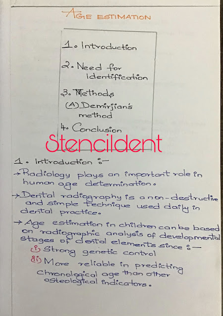
  AGE ESTIMATION  INTRODUCTION : Radiology plays an important role in human age determination Dental radiography is a non-destruction and simple technique used daily in dental practice  Age estimation in children can be based on radiographic analysis of developmental stages of dental elements since: Strong genetic control More reliable in predicting chronological age than other osteological indicators NEED FOR IDENTIFICATION: A competent forensic death investigation of human remains has 4 goals : Determination of the means Manner Cause of death Identification of the remains METHODS: DEMIRJIANS METHOD: To assess the developmental stages of third molars from the mandible . Eight stages (A-H) Plus 0 - absence  CONCLUSION : Teeth represent useful material for age estimation  Development of each individual can be effected by : Genetic  Nutritional Climatic  Hormonal Environmental  It has been reported that dental mineralisation is less affected by external factors when compared to bone mine

LEUKOPLAKIA - oral medicine notes

Image
 LEUKOPLAKIA  POTENTIALLY MALIGNANT DISORDER :  R isk of malignancy being present in a lesion or condition either during the time of initial diagnosis or at future date  PRECANCEROUS LESION :  Benign morphological altered tissue in which cancer is more likely to develop than its normal counterpart : leukoplakia  Erythroplakia  Tobacco pouch keratosis  Palatal lesion in reverse smokers  PRECANCEROUS CONDITION :  Generalized state or a disease which can be associated with greater than  normal risk of cancer development  OSMF Lichen planus  Epidermolysis bullosa  LEUKOPLAKIA :  White plaque of questionable risk having excluded (other)known disease or disorder that carry no risk for cancer  PLAQUE-  Raised lesion that are greater than 1 cm in diameter ,they are essentially large papules  PAPULE:  Lesion raised above skin or mucosal surface that are smaller than  1 cm in diameter  WHO DEFINITION : Non scrapable white patch or plaque that cannot be characterized clinically or pathologically